How AI Detects Cavities in Dental X-Rays, Explained
Understanding how AI detects cavities in dental x-rays comes down to one idea: the software compares millions of pixel patterns against images where a cavity was confirmed, then highlights the spots that match. Your dentist sees a colored box drawn over a suspicious area on the screen, along with a confidence score. The dentist still decides what it means.
This article was reviewed and updated in September 2026 to reflect four new meta-analyses and the latest FDA clearances. Below you will find how the detection works step by step, what the peer-reviewed accuracy numbers say, which systems have FDA clearance, and the limits researchers keep flagging that marketing pages tend to skip.
How Does AI Detect Cavities in Dental X-Rays?
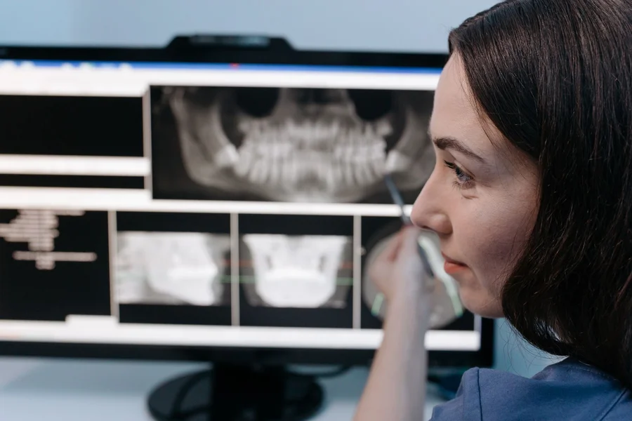
AI detects cavities in dental x-rays by scanning the image for density changes that match patterns it learned from thousands of labeled radiographs. A cavity shows up as a darker area where tooth mineral has been lost, and the software flags regions where that darkening matches known decay patterns rather than normal anatomy or image noise.
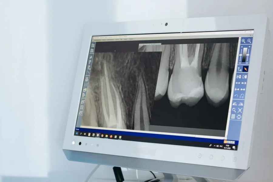
Two terms explain nearly all of it. A convolutional neural network, usually shortened to CNN, is a type of AI built specifically to read images, learning shapes and textures layer by layer rather than looking at a picture all at once. A bitewing radiograph is the standard side-view x-ray dentists take to see between back teeth, which is exactly where decay hides from a visual exam. The CNN does the pattern reading. The bitewing supplies the view that makes contact-point decay visible at all.
Practices adopting this software often pair it with other front-office AI, a shift covered in our piece on AI for small dental clinic scheduling. The workflow in a modern practice runs in three steps. The x-ray is captured and lands in the practice software. The AI analyzes it within seconds and draws a box around any suspected lesion, often with a percentage confidence attached. The dentist reviews the flagged areas alongside the unmarked image and makes the call. Regulators describe this as a “concurrent read” setup, meaning the software assists a clinician who is looking at the same image, rather than reading it alone.
How Accurate Is It Compared to a Dentist?
Pooled sensitivity sits between 0.76 and 0.94 depending on which review you read, with specificity between 0.85 and 0.94. That is generally at or above an unaided dentist reading the same image. Higher sensitivity means it catches more, not that it is always right.
Four separate meta-analyses published in 2026 have now measured this, and the honest summary is that the range is wide. Sensitivity means how often it correctly spots a cavity that is genuinely there. Specificity means how often it correctly leaves a healthy tooth alone.
- Umbrella review, Journal of Dentistry, searches to March 2026. Seventeen systematic reviews, five with pooled accuracy data. Sensitivity 0.76 to 0.94, specificity 0.85 to 0.91.
- BMC Oral Health, February 2026. Twenty-five studies, thirteen pooled. Sensitivity 0.86, specificity 0.91, AUC 0.94. Models reading intraoral photographs scored higher on sensitivity at 0.88, while models reading radiographs scored higher on specificity at 0.92.
- Dentomaxillofacial Radiology, July 2026. Proximal caries across bitewing, panoramic and periapical images. Sensitivity 76%, specificity 94%, AUC 0.90.
- Journal of Evidence-Based Dental Practice, March 2026. Proximal caries, twenty-eight studies. Sensitivity 0.84, specificity 0.93, and notably higher sensitivity on cone beam CT than on standard radiographs.
An earlier version of this article reported that decay between adjacent teeth was the strongest case for AI, with sensitivity around 0.94. The 2026 evidence points the other way. Both of the new proximal-caries meta-analyses put sensitivity at 0.76 and 0.84, at the bottom of the measured range rather than the top, while specificity holds up at 0.93 to 0.94. Read plainly, that means current software is better at leaving healthy contact points alone than at catching every lesion hiding between teeth. If you were told AI almost never misses decay between teeth, that claim no longer matches the pooled data.
The Dentomaxillofacial Radiology team also reported the raw spread before pooling, and it is the most useful number in any of these papers: individual model accuracy ranged from 28.5% to 100%. Averages hide that. “AI detects cavities” describes a category of software whose members perform wildly differently.
From what we’ve seen in how these numbers get quoted, the sensitivity figure travels far more widely than the specificity figure, and that imbalance matters. A tool that flags aggressively will score well on sensitivity while generating false positives that a dentist then has to rule out one by one.
Which Cavity-Detection Systems Have FDA Clearance?
Several do, and the count keeps climbing. Searching the FDA’s own 510(k) database under the two product codes these tools clear through returns roughly a dozen dental clearances between January and early September 2026 alone, from Overjet, Dentsply Sirona, Velmeni, Diagnocat, Orca Dental AI, Relu and Better Diagnostics AI.
The most recent caries-specific clearance at the time of writing is Overjet Multi-Image Caries and Charting Assist, K261059, cleared on 6 August 2026. Other recent entries worth knowing:
- Pearl Second Opinion Panoramic, K250525, decision date 14 November 2025, extending detection of caries, periapical radiolucencies and impacted third molars to panoramic images rather than bitewings alone.
- Videa Dental AI, K251002, decision date 19 September 2025. VideaHealth’s earlier caries product, Videa Caries Assist, cleared as K213795 back in April 2022.
- Overjet CBCT Assist, K251514, December 2025, and Overjet Iris, K253930, April 2026.
Two cautions about those dates. Company press releases often quote the announcement date rather than the FDA decision date, so the two can differ by a few weeks. And FDA’s database groups dental tools with all other radiological AI, so any headline count of “dental AI clearances” depends on how the person counting drew the boundary.
It helps to know what clearance means. These products go through the FDA’s 510(k) pathway, which establishes that a device performs comparably to something already on the market. It is a different and lighter standard than the full premarket approval used for high-risk medical devices. The review of FDA-cleared dental AI applications makes this point directly, noting that clearance verifies a system meets internal performance thresholds without guaranteeing consistent behavior across different clinical settings.
The same clearance-versus-evidence gap appears across diagnostic software generally, as our look at AI tools for computer diagnostics shows in a non-medical setting. Vendors publish their own figures too. Overjet and its competitors cite internal validation results on their sites, and those numbers are typically higher than what independent reviews find. VideaHealth, for instance, reports that in its FDA trial dentists using the software missed 43% fewer caries and made 15% fewer erroneous detections. That is a company-reported figure from the submission behind a 2022 clearance, not an independent replication. Treat company-published accuracy claims as a starting point rather than as verified performance.
What Changed in 2026?
Clearances sped up while the published evidence grew more cautious. That is the single most important development of the past year, and the two trends point in opposite directions.
On the regulatory side, dental AI approvals have accelerated every year since 2021, and 2026 has already added a dozen more. On the evidence side, the three largest 2026 reviews all landed on some version of the same verdict, and it is more guarded than what reviewers were writing in 2025.
Although AI models demonstrate high diagnostic performance under experimental conditions, current evidence does not support their use as stand-alone diagnostic tools.
That is the conclusion of the 2026 umbrella review in the Journal of Dentistry, which recommends implementation stay restricted to decision-support until prospective validation shows a real effect on clinical decisions and patient outcomes. The Journal of Evidence-Based Dental Practice meta-analysis graded its certainty of evidence as low and found 22 of its 28 included studies carried a high risk of bias. The BMC Oral Health authors recorded heterogeneity above 90%, meaning the studies disagreed with each other far more than chance explains.
None of that says the software does not work. It says the published proof is thinner than the approval count suggests, and that a cleared, widely sold product can still be waiting on the study that would confirm it changes outcomes. For a patient in the chair, the takeaway is unchanged but better supported than it was a year ago: the flag is a prompt for the dentist, not a verdict.
What Do Researchers Say the Limits Are?
Three limits come up repeatedly in the literature, and none of them appear on a product brochure. They concern who the training data represented, whether clearance predicts everyday clinical behavior, and what happens after deployment.
The first is selection bias in the studies themselves. The PLOS One authors noted that caries prevalence across the primary studies they analyzed was 27.3%, noticeably lower than prevalence in the general community. Accuracy measured on a cleaner-than-average sample of images does not automatically hold up on a typical day of patients.
The second is population coverage. The FDA-clearance review warns that when training datasets underrepresent certain age groups, ethnicities, anatomical variations, or imaging equipment, performance can vary across those populations. A model trained largely on one sensor brand may behave differently on another.
The third is the absence of ongoing monitoring. That same review flags the lack of systematic postmarket surveillance, meaning problems like model drift, where performance quietly degrades over time, may go undetected. Only two of the platforms reviewed had substantial independent published research behind them.
A fourth limit came into focus in 2026: the quality of the studies themselves. When the Journal of Evidence-Based Dental Practice team applied a formal risk-of-bias tool, 22 of 28 studies scored high risk, and the certainty of evidence came out low. Reviewers repeatedly flag the same causes, namely retrospective datasets, little external validation on images from other clinics, and inconsistent reporting of what counted as a cavity in the first place. A model tested only on the dataset it grew up with is being graded on homework it has already seen.
The myth worth clearing up: a flagged box on the screen is not a diagnosis, and it is not a treatment recommendation. The PLOS One authors state plainly that AI should not substitute for human judgment. A highlighted area means the software found a pattern worth a second look, and a dentist still has to weigh it against your clinical exam, your history, and the lesion depth.
What Should You Ask Your Dentist About It?
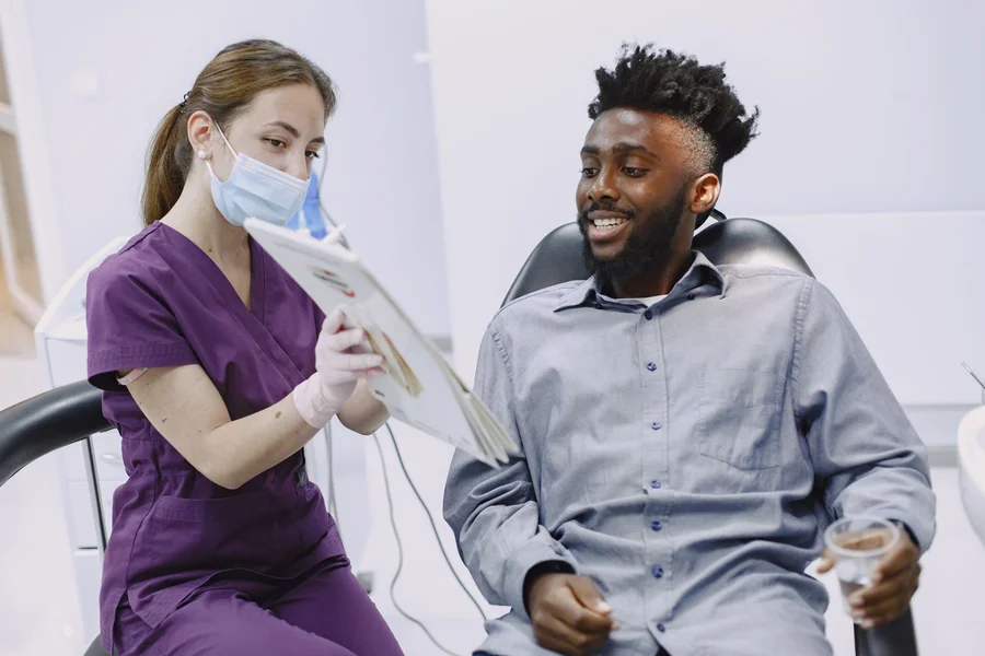
Ask what the software found, how confident it was, and what the alternative to treating right now would be. A dentist using these tools well should be able to answer all three without hesitation.
- “Can you show me what the AI flagged?” Most systems display the marked image on screen. Seeing it yourself makes the conversation concrete.
- “Do you agree with the finding?” The dentist’s own read is the one that matters, and a good clinician will say when they disagree with a flag.
- “How far into the tooth does it go?” Decay confined to enamel is often monitored rather than drilled, while decay reaching dentin usually needs treatment.
- “What happens if we watch it instead?” Monitoring is a legitimate option for early lesions, and the answer tells you how urgent the finding really is.
- “Was this visible before the AI flagged it?” This distinguishes a genuine catch from a borderline call the software surfaced.
A common mistake worth avoiding: treating a high confidence percentage as certainty about whether you need a filling. A 90% confidence score describes the software’s pattern match on an image, not the clinical necessity of drilling your tooth. Those are different questions, and only the second one determines your treatment. If a treatment plan expands substantially right after a practice adopts new detection software, it is reasonable to ask for a second opinion.
After looking at how these tools get discussed with patients, we prefer practices that show you the unmarked image alongside the flagged one. Seeing both makes it obvious whether the finding is a clear dark shadow or a subtle judgment call, and that context is genuinely useful when you are deciding about treatment.
The same assist-not-replace pattern shows up wherever image-reading AI has been deployed, as our piece on AI vision systems for vehicle inspections describes in a lower-stakes setting.
Frequently Asked Questions
Does AI cavity detection mean more radiation exposure?
No. The AI analyzes x-rays that were already taken as part of normal care and does not require additional or stronger images. If a practice suggests extra x-rays specifically to feed the software, ask why, since the standard imaging schedule is based on your individual risk, not on software requirements.
Can AI find cavities that a dentist would miss?
Sometimes, particularly early decay between teeth where the visual signal is faint. Studies comparing AI to unaided dentists consistently show higher average sensitivity for the software. The tradeoff is that it also flags areas that turn out not to be decay, which is why a clinician reviews every finding.
What is the difference between AI detection and a regular dental x-ray reading?
A regular reading depends entirely on the dentist’s eye and experience with that single image. AI detection adds a second pass that compares the image against patterns learned from thousands of confirmed cases, then marks candidates for the dentist to evaluate. The x-ray itself is identical either way.
Is AI cavity detection approved by the FDA?
Several products have FDA clearance through the 510(k) pathway, with 29 cleared dental imaging products from 13 companies counted as of July 2025. Clearance is not the same as full premarket approval, and it demonstrates comparability to existing devices rather than proving superior patient outcomes.
Should I get a second opinion if AI flags a cavity?
A second opinion is reasonable any time a treatment plan feels larger than expected, whether AI was involved or not. Ask your dentist to show you the image and explain the lesion depth first, since that conversation often resolves the question without another appointment.
Does AI work on all types of dental x-rays?
Performance varies by image type, and most validated caries research focuses on bitewing radiographs. Detection on panoramic images and 3D cone beam scans is a separate capability with its own clearances and its own accuracy figures, so results from bitewing studies do not transfer automatically.
Has AI cavity detection got more accurate over the last year?
The measured numbers have not improved, and for decay between teeth they came down. Two 2026 meta-analyses put proximal caries sensitivity at 0.76 and 0.84, below the 0.94 that narrower earlier reviews reported. What did improve is the quality of the measurement, since 2026 brought larger reviews that pooled more studies and judged them more strictly.
If the FDA cleared it, does that mean it has been proven to work?
No. A 510(k) clearance shows a device is comparable to something already on the market, which is a lighter standard than proving clinical benefit. The 2026 umbrella review in the Journal of Dentistry concluded that current evidence does not support these tools as stand-alone diagnostics, even though dozens of them are cleared and selling. Clearance and proof of outcome are separate questions.
Will AI replace dentists reading x-rays?
Not based on how these systems are currently cleared or how researchers describe them. The FDA clearances cover assistive concurrent-read use, meaning a clinician reviews the same image, and the PLOS One review authors explicitly state AI should not substitute for human judgment.
What to Take Away About How AI Detects Cavities in Dental X-Rays
How AI detects cavities in dental x-rays is genuinely useful technology sitting behind a screen at a growing number of practices, with pooled sensitivity around 0.85 and specificity around 0.90 in the best available synthesis of the research. It catches early decay that is easy to miss, and it flags things that turn out to be nothing.
If your dentist uses one of these systems, ask to see the flagged image at your next visit and ask about the lesion depth before agreeing to treatment. That single question turns a highlighted box on a screen into a conversation you can genuinely take part in.
This article is general information about how the technology works, not dental or medical advice. Decisions about your own treatment should be made with your dentist, who can assess your specific clinical situation.